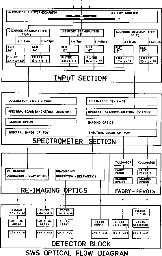
SWS has three different apertures (aperture 4, used by the LW FP, is a virtual aperture offset slightly from aperture 3 to increase the amount of light going to the FPs and hence improve efficiency). A shutter system allows the selection of one aperture, while closing off the other two (the spacecraft pointing has to be adjusted so that the target is imaged onto the selected aperture).
Each aperture is used for two wavelength ranges, one for the short-wavelength (SW) section of the spectrometer and one for the long-wavelength (LW) section. Since those two sections are otherwise independent, two wavelength ranges can be observed simultaneously .
Beamsplitters , consisting of
Reststrahlen crystal filters ( ![]() , LiF and
, LiF and ![]() ), are located
behind the apertures. The beams
transmitted by the first crystal enter the SW section; the reflected
beams enter the LW section, after a second reflection against identical
material. As is seen in the schematic (Fig. 2.3), the
actual entrance slits are located behind the beam-splitting crystal. In
this way, each of the 6 possible input beams has its own slit. All
slits have been given the same width, except for the
), are located
behind the apertures. The beams
transmitted by the first crystal enter the SW section; the reflected
beams enter the LW section, after a second reflection against identical
material. As is seen in the schematic (Fig. 2.3), the
actual entrance slits are located behind the beam-splitting crystal. In
this way, each of the 6 possible input beams has its own slit. All
slits have been given the same width, except for the ![]() reflected
input, which has a larger width, adapted to the larger diffraction image
at these wavelengths (see Tab. 2.1). The slits are in the
focus of the telescope, in the plane where the sky is imaged.
reflected
input, which has a larger width, adapted to the larger diffraction image
at these wavelengths (see Tab. 2.1). The slits are in the
focus of the telescope, in the plane where the sky is imaged.
In the direction perpendicular to the dispersion, the slits are oversized. There the fields-of-view are determined by the dimensions of the detectors. The cross-dispersion dimensions are different for almost all detector bands. Since the imaging of the slits onto the detectors (or vice versa) is imperfect due to aberrations and diffraction , the short sides of the fields of view (detector edges) are more fuzzy than the long sides (the slit jaws).
Small offsets of the fields of view perpendicular to the dispersion can be caused by alignment errors. The internal alignment specification adhered to amounts to 10% of the detector size alias spectrum width.
The monochromatic images of the grating detectors fill about 55% of the slit
widths. This means that the spectral resolution for point sources is
significantly higher than for extended sources, in a ratio that is affected, of
course, by diffraction (see Fig 5.2,
p.  ). The resolution figures in this document apply to
extended sources only.
For the Fabry-Pérot section the situation is more complex. There the monochromatic detector images just fill the slit width. With the spectral channeling by the F-Ps, a subtle interplay arises between spectral and spatial properties. Effectively, the spatial resolution may increase due to the narrowness of the F-P resonance. Point sources have less leakage in unwanted F-P orders than extended sources. Spatial extent does not influence the spectral resolution of the F-Ps.

Figure 2.2: Block diagram of the SWS. The diagram shows the optical
functions of the spectrometer, excluding its internal calibration
sources, but including the shutter, the collimation and the imaging
optics. It excludes band 3E.

Figure 2.3: Optical schematic of the SWS.
The diagram indicates the location of the six separate entrance slits
behind the dichroics after the three apertures. It shows all the
spectral order-separation filters and the internal wavelength
calibrators. The shutters, collimation and imaging optics and band 3E have
been left out